CT
CT and ZP
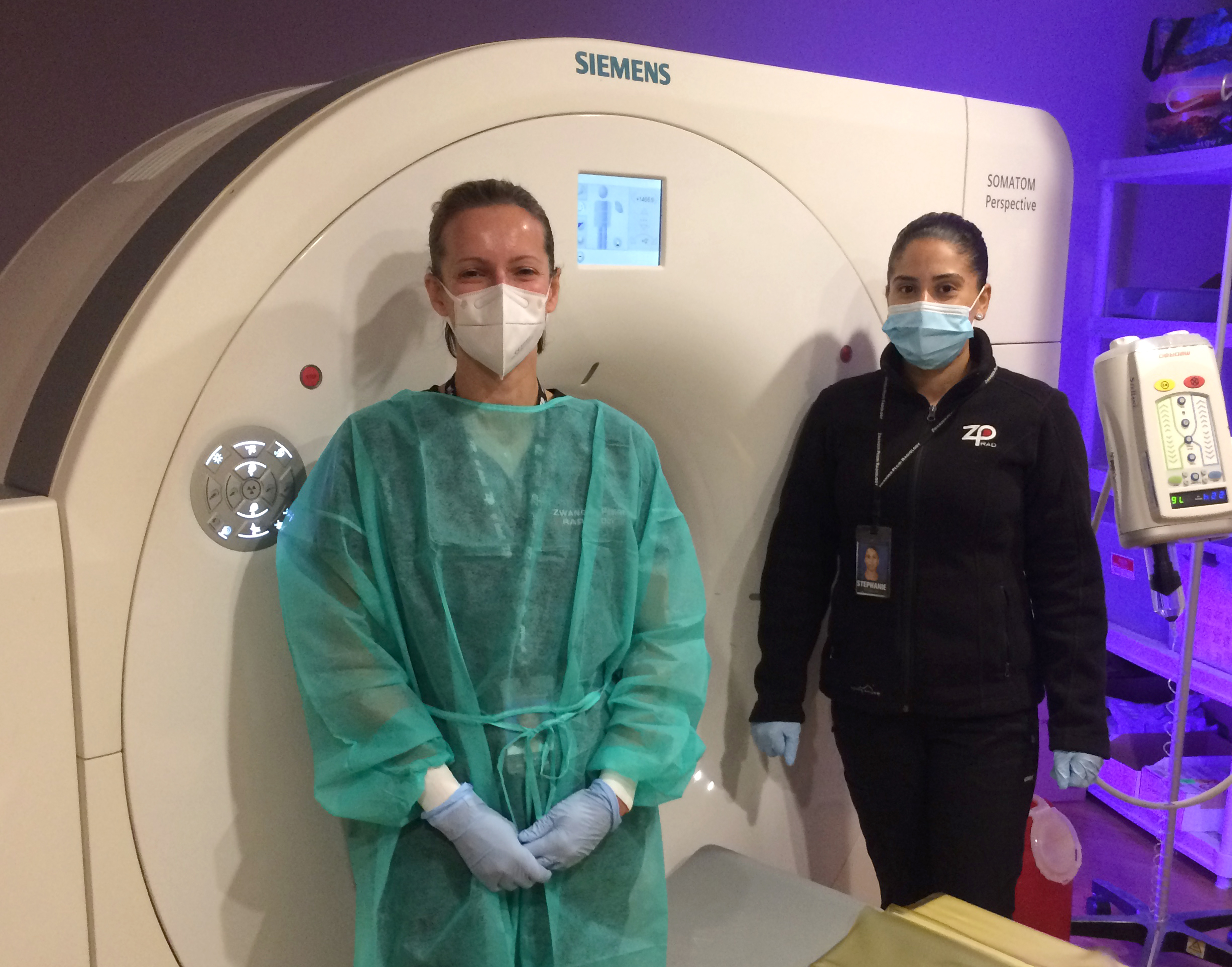
Over the past several years, we have been dedicated to overhauling our entire inventory of CT (computed tomography) scanners and replacing all machines with the best equipment available. This guarantees patients will be exposed to the smallest amount of radiation possible while still getting high-quality images.
ZP follows three principles which promise patient safety and low doses of radiation: ALARA (As Low As Reasonably Achievable), Image Wisely™, and Image Gently. Our commitment to these principles is reflected by the patent awarded to Dr. Steven Mendelsohn, CEO of Zwanger-Pesiri Radiology, in 2012, for a CT dose card (US Patent #: US8,281,996B2). This card was given to every patient after their scan to inform patients of the radiation dose they were exposed to during their test. Now this number is generated on every report so doctors and patients are kept informed.

How CT Works
CT scans use X-rays taken from multiple angles as a patient is moved through the opening of the machine. An X-ray tube and high resolution digital detector rotate very fast inside the machine’s opening to obtain pictures from all different angles and produce detailed images of bones, soft tissue, organs, and blood vessels. The rotation of these parts is internal and cannot be detected by the patient. The images produced from a CT scan are significantly more detailed than a traditional X-ray. It is considered an essential tool to assess acute brain trauma, kidney stones, and other conditions.
The CT scanner takes many very thin two-dimensional pictures, which the computer can assemble into three-dimensional images. This allows the doctor to look layer by layer at the area being scanned and provides greater detail to aid in the diagnostic process.
Many CT scans require the use of a contrast dye. The contrast may be given in a drink that you consume prior to the scan or administered during the scan through an I.V. The contrast highlights certain parts of your body and helps to provide the sharpest images available.


Benefits of a CT Examination
CT can image bone, soft tissue, and blood vessels at the same time
CT scanning is painless, noninvasive, and accurate
CT imaging provides real-time imaging
CT scanners produce superior exams using a fraction of time and radiation exposure
Why Choose Zwanger Pesiri?
Zwanger-Pesiri Radiology brings world-class expertise to the Long Island community. Our subspecialty-trained radiologists are Board Certified by the American Board of Radiology with fellowship training in a variety of specialties. They are highly-skilled, highly-knowledgeable, and make patient care a priority. To learn more, contact us today.
